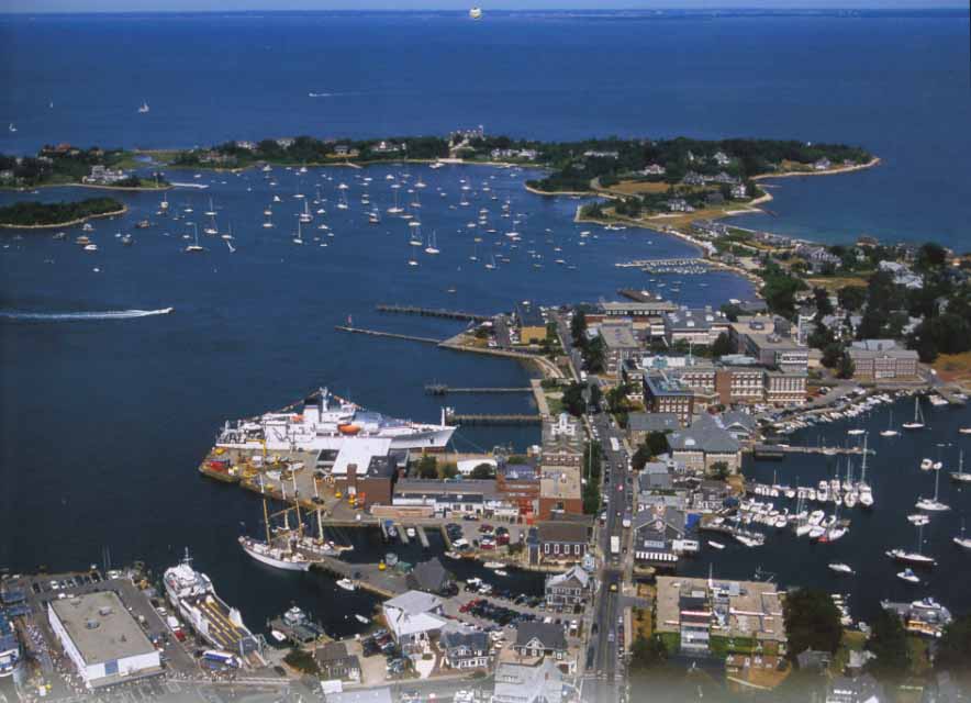
Andrew attended the 2017 Woods Hole Optical Microscopy and Imaging in the Biomedical Sciences course in Woods Hole, MA from September 6-16th. Andrew was selected to be one of only 25 students to attend this high-level microscopy course! Topics to be covered include: (a) fundamental principles of microscope design, image formation, resolution, contrast; (b) bright field, dark field, phase contrast, polarized light, differential interference contrast, interference reflection, and fluorescence microscopy; (c) cameras, signal to noise ratio, digital image recording, processing and analysis, multispectral imaging; (d) advanced fluorescence– fluorescent probes, TIRF, FRET, FLIM, FRAP, polarization of fluorescence, fluorescence correlation spectroscopy; (e) digital image restoration/deconvolution, and 3-D imaging principles, confocal scanning microscopy, multiphoton excitation fluorescence microscopy, light-sheet microscopy; application of the optical methods to live cells will be emphasized, although other specimens also will be discussed; (f) super-resolution techniques including localization microscopy, stimulated emission depletion microscopy (STED), structured illumination microscopy. Particular emphasis will be placed on ‘picking the right tool for the job’. Congrats, Andrew!

 Andrew and Paul publish a new paper with Dr. Chris Ross (Johns Hopkins)


